
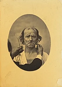
Plate 14: Designed to show a comparison between the maximal contraction of the superior part of m. orbicularis oculi and m. corrugator supercilii, in the same individual. On the left, very strong electrization of the superior part of m. orbicularis oculi: dissatisfaction, somber thoughts. On the right, electrization of m. corrugator supercilii.
1854-1856, printed 1862
Medium
albumen print
Dimensions
image: 22.1 × 15.9 cm (8 11/16 × 6 1/4 in.) mount: 40.4 × 27.7 cm (15 7/8 × 10 7/8 in.)
Classification
Photograph
Department
CPH
Museum
National Gallery of Art, Washington, D.C.
Credit
Eugene L. and Marie-Louise Garbáty Fund
Accession Number
2015.52.19
Rights
Public Domain
About the Artist
Guillaume-Benjamin-Amant Duchenne (de Boulogne)
Guillaume-Benjamin-Amant Duchenne de Boulogne (1806–1875) was a French neurologist whose pioneering use of photography to document human facial expression placed him at the unique intersection of medical science and visual art. Though primarily a physician rather than an artist in the conventional sense, his photographic work — produced in the 1850s and 1860s — has been recognized as a significant...